Posted by on July 14, 2010 - 12:05pm

CHICAGO --- The more an older woman weighs, the worse her memory, according to new research from Northwestern Medicine. The effect is more pronounced in women who carry excess weight around their hips, known as pear shapes, than women who carry it around their waists, called apple shapes.  The study of 8,745 cognitively normal, post-menopausal women ages 65 to 79 from the Women's Health Initiative hormone trials is the first in the United States to link obesity to poorer memory and brain function in women and to identify the body-shape connection.

"The message is obesity and a higher Body Mass Index (BMI) are not good for your cognition and your memory," said lead author Diana Kerwin M.D., an assistant professor of medicine and a physician at Northwestern Medicine. "While the women's scores were still in the normal range, the added weight definitely had a detrimental effect."

For every one-point increase in a woman's BMI, her memory score dropped by one point. The women were scored on a 100-point memory test, called the Modified Mini-Mental Status Examination. The study controlled for such variables as diabetes, heart disease and stroke.   The study will be published July 14 in the Journal of the American Geriatric Society.

The reason pear-shaped women experienced more memory and brain function deterioration than apple-shaped women is likely related to the type of fat deposited around the hips versus the waist. "Obesity is bad, but its effects are worse depending on where the fat is located," Kerwin said.

Cytokines, hormones released by the predominant kind of fat in the body that can cause inflammation, likely affect cognition, Kerwin said. Scientists already know different kinds of fat release different cytokines and have different effects on insulin resistance, lipids and blood pressure.

"We need to find out if one kind of fat is more detrimental than the other, and how it affects brain function," she said. "The fat may contribute to the formation of plaques associated with Alzheimer's disease or a restricted blood flow to the brain."    In the meantime, the new findings provide guidance to physicians with overweight, older female patients.

"The study tells us if we have a woman in our office, and we know from her waist-to-hip ratio that she's carrying excess fat on her hips, we might be more aggressive with weight loss," Kerwin said. "We can't change where your fat is located, but having less of it is better."

Kerwin's research is funded by the T. Franklin Williams Award from Atlantic Philanthropies and Association of Specialty Professors and the Wisconsin Women's Health Foundation Faculty Scholar Award. The Women's Health Initiative was funded by a grant from the National Heart, Lung and Blood Institute.

Marla Paul is the health sciences editor. Contact her at marla-paul@northwestern.edu

Posted by on July 13, 2010 - 4:11pm

A recent post on the Oncofertility Consortium Blog discussed gender disparities in the senior levels of scientific research. Women receive 56% of science and engineering undergraduate degrees and are awarded more than 40% of graduate degrees in the sciences, often a PhD. However, they make up only 22% of senior academic faculty members in the United States.

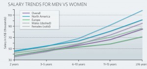
The Journal Nature may have come across another reason for the gender gap in science. Salary differences. Nature just released the results of their first-ever salary and career survey of more than 10,000 scientists. In addition to examining salaries across countries, academic stages, and industry, the study also looked across genders.

The report found that female scientists begin their post-graduate careers making slightly more than male scientists, about $45,000 per year in the United States. However, 5 years after receiving their highest degree, when scientists generally begin their first academic appointments, male scientists start to outpace females. As time progresses, this trend continues so that 16 or more years past degree completion, men make about $120,000 while female scientists hover below $105,000.

It is important to note that similar salary trends occur in both North America and Europe. According to the study, “Men’s salaries were 18% to 40% higher than women’s in the countries for which we had significant sample sizes-Australia, Germany, Italy, Spain, the United Kingdom, India, Japan, Canada, and the United States.”

The exact cause of the scientific wage gap is unknown. However, in my previous career as a scientist, I personally saw women poorly negotiate for starting salaries, producing an initial wage difference that increased over time. In addition, some of my fellow female scientists either took time off from work to raise children or opted for more-flexible, lower-paying, non-tenured positions. In my case, which occurs with many women, I foresaw that my significant other would make more money the long-term and saw myself sacrificing my career for our future family. In my transition away from the bench, I have instead avoided the “sex, science, and salary” issue altogether but the scientific community needs to learn how to keep women in the sciences or risk future scientific and medical advances. The most obvious way to do that? Money.

--

Kate is on loan from the Oncofertility Consortium. Check out their blog!

Posted by on July 13, 2010 - 3:27pm

Fish and shellfish are an important part of a healthy diet.  They contain protein and other essential nutrients,  are low in saturated fat, and contain omega-3 fatty acids.  A well-balanced diet that includes a variety of seafood can contribute to heart health and children’s proper growth and development.

However, nearly all seafood contains traces of mercury.   For most people, the risk from mercury in fish is not a health concern.   Yet, some fish and shellfish contain higher levels of mercury that may harm an unborn baby or young child’s developing nervous system and this depends on the amount of seafood eaten and the levels of mercury in these foods.

The Food and Drug Administration (FDA) and the Environmental Protection Agency (EPA) advise that women who may become pregnant, pregnant women, nursing mothers, and young children avoid some types of fish and shellfish.

To reduce exposure to mercury while obtaining benefits of eating fish, the FDA recommends that you:

1.   Do not eat shark, swordfish, king mackerel or tilefish because they contain high levels of mercury.

2.  Eat up to 12 ounces (2 average meals) a week of a variety of fish and shellfish that are lower in mercury (shrimp, canned light tuna, salmon, Pollock, and catfish).

What effect does the oil spill have on the fish in the Gulf of Mexico?

The FDA is working with the National Oceanic and Atmospheric Administration (NOAA) National Marine Fisheries Service, the EPA, other Federal agencies and several state authorities in the regions affected by the recent oil spill.  Federal and state officials are monitoring the waters from which seafood is harvested and will act to close areas contaminated by the oil spill to fishing.  A large area of the Gulf of Mexico and been closed to commercial fishing and the FDA has authority to seize any adulterated seafood that may show up in markets.

According to the FDA:

  • Although crude oil has the potential to taint seafood with flavors and odors cause by exposure to hydrocarbon chemicals, the public should not be concerned about the safety of seafood in stores at this time.
  • Fish and shellfish harvested from areas unaffected by the closures are considered safe to eat.

For more information about the effect of the oil spill on pregnant women, click here

You can call 1-888-INFO-FDA if you have concerns about seafood you have purchased that you suspect is contaminated with oil.

Posted by on July 12, 2010 - 9:41am

L-R:    Shaquita, Dr. Carla Pugh, Megan,  and Nicole

The Oncofertility Summer Research Fellowship 2010 is now in full swing (check out this post for more details)!  Our three Oncofertility Saturday Academy alumni undergraduate students, Nicole Miles, Shaquita Webster, and Megan Romero have been hard at work, both at the bench in Dr. Woodruff's laboratory and learning about many social issues surrounding women's health and oncofertility.  Last week, Dr. Carla Pugh presented her work on the use of technology to improve clinical skills education at the IWHR Monthly Research Forum, and Shaquita, Megan, and Nicole wrote the following post on their thoughts about the event.  Enjoy!

On June 22, 2010 at the Institute for Women’s Health Research Monthly Research Forum,  Carla Pugh, MD,  Assistant Professor of Surgery and Associate Director of the Center for Advanced Surgical Education here at Northwestern University, spoke about some of the anxiety and concerns first year medical students face when performing pelvic, prostate, and breast exams.  She stated that the major reason many medical students are so apprehensive to administer these ‘intimate’ exams is due to their lack of experience, as many are fresh out of college. As a first year medical student they are bombarded with information on the anatomy of the human body. However, in order to become a good physician one must not only have an understanding of the human anatomy but also how to perform a physical examination.

With this knowledge Dr. Pugh developed numerous pelvic, prostate, and breast anatomic models linked to sensors. These sensors are linked to computers that display where and how much pressure is applied to various anatomic features of the pelvic, prostate, and breast models.

Dr. Pugh demonstrated that the leading cause of anxiety for second year med students was fear of missing a lesion. Due to breast cancer’s prominence in women, the fear of missing something during a clinical breast exam can be a particularly anxiety-causing. Dr. Pugh also commented on the differences between the ways that clinical breast exams are performed by male and female practitioners, in terms of the time spent and amount of pressure used during the exam.

What makes the mannequins so amazing is that the sensors are placed inside model’s vagina or rectum, and as students' fingers move along the anatomy, they can see the location of their hands on a screen. "When you put your finger in someone's rectum for the first time, you think you know where the prostate is, but you don't," Pugh said. "Several things in medicine are assumed to be learned in time, but I think we can do better than that. You can't teach everybody everything in a lecture format."

Dr. Pugh’s inventions have revolutionized the world of medicine in various ways. But there was one thing that she had not mentioned.  After the seminar, we asked Dr. Pugh how her models are used outside of the classroom and whether they are available to local community centers so that women who don’t have the resources to get to a doctor can visit the center and teach themselves how to perform the exam.  We believe the breast models, in particular, would be beneficial to individuals in the community.  If women are able to give exams on the models it will help encourage self breast exams and promote early detection. While she was amazed by such a great thought, her response was that she had not had the opportunity to do so.

After a discussion with Dr. Pugh, we were interested in finding a way to get the breast  models out into the community.  Throughout our fellowship in the Woodruff lab, we are beginning to understand that the best way to learn any concept is by teaching it to someone else. We suggested creating a community outreach portion for her medical students.  Unfortunately, Dr. Pugh has no control over the curriculum nor does she have enough time with the students to implement such a plan. However, she stated that she would love to have our help to make it possible. With our mentors as our witness, taking Dr. Pugh's breast models to community centers across the city in efforts to increase early detection of breast cancer is now a goal in the making!

Dr. Pugh is also featured in an article in this week's Chicago Tribune.

Authors:  Megan Romero, Nicole Miles, and Shaquita Webster (Oncofertility Summer Research Fellows)

Posted by on July 9, 2010 - 11:37am

In March 2010, Congress passed and President Obama signed the Affordable Care Act--the new health care reform law.  It will take several years for all provisions of the Act to be implemented but there are a variety of tools becoming available to help guide the way as provisions are implements.   The Our Bodies, Ourselves Blog has posted one such tool.   To access it, click here

Posted by on July 8, 2010 - 11:18am

Several Northwestern researchers, including our own Institute director, Teresa K. Woodruff, PhD, have been making a strong case for more sex-based research that is making waves in several prestigious journals including Nature and Women's Health.   Readers interested in reading these articles should click here

Posted by on July 7, 2010 - 4:50pm

Many people assume that brain differences between men and women result from sex hormones like estrogen and testosterone. This is not true. In fact, increasing numbers of studies find large reliable distinctions between the sexes. These effects are often seen early in development and before sex hormones are expressed. The research examining sex-specific brain mechanisms finds variability in structure, neurotransmitters, and the expression of specific genes in males and females.

Sex differences in size, shape, and function occur in every region of the brain. For example, sex divergence is seen in the hippocampus, an area of the brain that is important for learning and memory. The hippocampus makes up a larger portion of total brain size in women than in men. But that doesn’t necessarily mean that women learn better than men. Interestingly, some sub-regions of the hippocampus are larger and contain more cells in men. Further studies will be needed to fully understand the implications of such sex differences.

Additional variation occurs in the levels of neurotransmitters, the chemical messengers, in the brain. Males and females differentially produce serotonin, a neurotransmitter that affects mood. These differences may have significant effects on gender-specific treatment of diseases, such as depression.

Gene expression differences between the sexes are also seen across many species. Scientists have found groups of genes with sex-specific expression that are conserved, or maintained, across primate species, suggesting that such patterns are inherited across evolution.

What is the evolutionary advantage to having sex differences in the brain? While we can only guess, one hypothesis is that such variability is important in the selection animals make in finding mates and raising offspring. Whatever the cause, differences in the brain affect development and health during adulthood. Many of the genes with differential expression are also associated with neurological diseases such as Alzheimer’s disease, schizophrenia, and cerebral ischemia. As such, sex-specific treatments of these diseases should also be studied.

__________________________

Kate is also a frequent contributor to the Oncofertility Consortium Blog. Check it out!

Posted by on July 6, 2010 - 10:28am

A survey of more than 1,200 primary care physicians indicates that many are not following clinical practice guidelines on recommended screening intervals for cervical cancer, both with regard to traditional Pap testing as well as a newer screening method, a DNA test for the human papillomavirus (HPV). The FDA has approved HPV DNA testing for use in conjunction with Pap testing, a process called co-testing, for women age 30 and older.

At the time the survey was conducted, guidelines from the American Cancer Society and those from the American Congress of Obstetricians and Gynecologists advised extending the interval between screenings to 3 years for low-risk women over the age of 30 after three consecutive normal Pap tests or a single normal co-test (a normal Pap test plus a negative HPV DNA test). Guidelines from the U.S. Preventive Services Task Force also are consistent with a longer interval between screening tests.

In the survey, based on a hypothetical clinical vignette of a 35-year-old, low-risk woman with three prior normal Pap tests, only 32 percent of respondents reported that they would comply with guideline recommendations, researchers from the CDC and NCI reported in the June 14 Archives of Internal Medicine. Even fewer respondents, 19 percent, would comply when, during a single visit, the low-risk woman had a normal co-test result. Approximately 60 percent of those surveyed—which included general internal medicine physicians, family practice doctors, and obstetrician-gynecologists—said they would still recommend that the woman undergo annual Pap screening.

Although the Pap test is the most commonly used cervical cancer screening method, a number of studies have shown that the DNA test for HPV—the cause of the vast majority of cervical cancer cases—is more sensitive than the Pap test in detecting high-grade precancerous lesions, spurring discusions about the optimal approach to cervical cancer screening in the United States.

This new study, however, suggests that guidelines for extending screening intervals have not influenced current clinical practice, wrote Dr. Mona Saraiya of the CDC’s Division of Cancer Prevention and Control and her colleagues. “When offered the choice for HPV testing,” they wrote, “many physicians deferred to the same pattern they used for Pap testing,” annual screening with both tests or no recommendation for HPV testing.

“These practice patterns are not likely to lead to much improvement in cervical cancer outcomes, but may result in unnecessary follow-up testing, increased risk of colposcopy-associated morbidities, and distress for patients,” said Dr. Robin Yabroff, a study co-author from NCI’s Division of Cancer Control and Population Sciences.

“New HPV infections are extremely common but overwhelmingly benign; they almost always go away by themselves,” said Dr. Mark Schiffman of NCI’sDivision of Cancer Epidemiology and Genetics. “Only persistent infections are a risk factor for cancer. If you screen for HPV too often, you will detect new infections rather than persistent infections, and this poses the risk of overtreatment.”

Posted by on July 1, 2010 - 10:35am

Women who measure their peak heart rates for exercise will need to do some new math, as will physicians giving stress tests to patients.  A new formula based on a large study from Northwestern Medicine provides a more accurate estimate of the peak heart rate a healthy woman should attain during exercise. It also will more accurately predict the risk of heart-related death during a stress test.

“Now we know for the first time what is normal for women, and it’s a lower peak heart rate than for men,” said Martha Gulati, MD, assistant professor of medicine and preventive medicine and a cardiologist at Northwestern Medicine. “Using the standard formula, we were more likely to tell women they had a worse prognosis than they actually did.”   Gulati is the lead author of a study published June 28 in the journal Circulation.

“Women are not small men,” Gulati added. “There is a gender difference in exercise capacity a woman can achieve. Different physiologic responses can occur. ”   Gulati was the first to define the normal exercise capacity or fitness level for women in a 2005 study.

The old formula -- 220 minus age -- used for almost four decades, is based on studies of men. The new formula for women, based on the new research, is 206 minus 88 percent of age.   At age 50, the original formula gives a peak rate of 170 beats per minute for men and women. The new women’s formula gives a maximum heart rate of 162 beats for women.  Many men and women use their peak heart rate multiplied by 65 to 85 percent to determine their upper heart rate when exercising.

“Before, many women couldn’t meet their target heart rate,” Gulati said. “Now, with the new formula, they are actually meeting their age-defined heart rate.”    The new formula is trickier to calculate, Gulati acknowledged, but is easily determined with a calculator. She currently is working on an iPhone application for a quick calculation.

The new formula is based on a study of 5,437 healthy women ages 35 and older who participated in the St. James Women Take Heart Project, which began in the Chicago area in 1992.    With the new formula, physicians will more accurately determine if women are having a normal or abnormal response to exercise.    “If it’s abnormal, that’s a marker for a higher risk of death,” Gulati said. “Maybe we need to talk about whether you exercise enough and what we need to do to get it into the normal range.

“We need to keep studying women to get data applicable to women,” Gulati said. “It’s important to not get complacent that we have data on men and assume women must be the same. They’re not.”

Gulati’s senior author on the study was the late Morton Arnsdorf, MD, professor emeritus and associate vice chairman of medicine and former section chief of cardiology at the University of Chicago.

Posted by on June 25, 2010 - 9:14am

The following press release was issued by the FDA. While the seriousness and pain of bone fractures is significant, the side effect profiles of this drug sound pretty serious.   What is the risk/benefit ratio?   Is the trade-off worth it?  We would love to hear from women who are making these choices.

JUNE 1, 2010:   The U.S. Food and Drug Administration today approved Prolia, an injectable treatment for postmenopausal women with osteoporosis who are at high risk for fractures.

Osteoporosis is a disease in which the bones become weak and are more likely to break. According to the National Institute of Arthritis and Musculoskeletal and Skin Diseases, 80 percent of the people in the United States with osteoporosis are women. One out of every two women over age 50 will break a bone in their lifetime due to osteoporosis.

People with osteoporosis at high risk for fracture include those that have had an osteoporotic fracture, or have multiple risk factors for fracture; or those who have failed or are intolerant to other available osteoporosis therapy. Prolia works to decrease the destruction of bone and increase bone mass and strength. An injection of Prolia is recommended once every six months.

“Due to its prevalence, osteoporosis is a serious concern to public health,” said Julie Beitz, M.D., director of the FDA’s Office of Drug Evaluation III. “The approval of Prolia provides another treatment option for postmenopausal women with osteoporosis who are susceptible to fractures.”

The safety and efficacy of Prolia in the treatment of postmenopausal osteoporosis was demonstrated in a three-year, randomized, double-blind, placebo-controlled trial of 7,808 postmenopausal women ages 60 to 91 years. In the study, Prolia reduced the incidence of vertebral, non-vertebral, and hip fractures in postmenopausal women with osteoporosis.

The most common side effects reported with Prolia include back pain, pain in the extremities, musculoskeletal pain, high cholesterol levels, and urinary bladder infections. Serious adverse reactions include hypocalcaemia (low calcium levels in the blood), serious infections, including infections of the skin, and dermatologic reactions such as dermatitis, rashes, and eczema.

Prolia causes significant suppression of bone turnover and this suppression may contribute to the occurrence of osteonecrosis of the jaw, a severe bone disease that affects the jaw, atypical fractures, and delayed fracture healing.

Prolia was approved with a risk evaluation and mitigation strategy (REMS) that includes a Medication Guide for patients and communications to health care providers that explains the risks and benefits of the drug.

Prolia is manufactured by Amgen Manufacturing Limited, a subsidiary of Thousand Oaks, Calif.-based Amgen Inc.

For more information about osteoporosis click here.

Pages